


Application of Personalized 3D-Printed Aortic Root Models with Built-In Sensors in Minimally Invasive Surgery
Release date:
2025-12-25 17:52
This paper demonstrates the use of 3D printing to fabricate multi-material, patient-specific aortic root models incorporating an embedded sensor array. The validity of the models was evaluated by comparison with corresponding patient data.
Abstract
Minimally invasive surgery offers significant advantages; however, insufficient anatomical understanding of the treatment target area can readily lead to postoperative complications. Transcatheter aortic valve replacement (TAVR) represents a minimally invasive therapeutic approach for aortic stenosis. In this study, we propose a multi-material 3D-printing technique to fabricate patient-specific, flexible aortic root models embedded with an array of electronic sensors. The validity of these models is validated by comparing their geometric congruence with postoperative patient data and by assessing in vitro hemodynamic performance, both with and without leaflet calcification. The integrated sensors enable the mapping of pressure distributions across key anatomical regions of the aorta, thereby facilitating optimized selection of bioprosthetic valves and comprehensive ex vivo implantation evaluation. This approach paves new avenues for reducing the risk of postoperative complications and advancing the development of next-generation medical devices. Importantly, this technological direction is highly aligned with clinical translation needs. As a provider of high-end medical testing equipment and services, Xi’an Dewei Medical has leveraged its highly realistic simulation models and comprehensive testing solutions to play a pivotal role in the R&D and validation of minimally invasive interventional medical devices. 。
Introduction
Global population aging is accelerating, with the U.S. population aged 65 and older projected to reach 73.1 million by 2030. This demographic shift is driving up the prevalence of cardiovascular diseases; aortic stenosis (AS) is a common degenerative condition in older adults, with as many as 2.7 million patients aged 65 and older in North America alone. AS restricts valve motion, impeding blood flow and ultimately leading to ventricular dysfunction. Given the advanced age and comorbidities of these patients, some are at high risk for conventional open-heart valve replacement surgery. Transcatheter aortic valve replacement (TAVR), which involves delivering a bioprosthetic valve via a catheter to the site of the lesion, has emerged as the preferred treatment option for this patient population.
Postoperative complications of TAVR primarily include paravalvular leakage and conduction disturbances, which are closely related to individual patient anatomical characteristics, the surgical technique employed, and the properties of the prosthetic valve itself. Optimizing these factors based on patient anatomy is crucial for preoperative planning and for reducing the risk of complications. Unlike conventional open-heart surgery, the minimally invasive nature of TAVR makes it challenging for the operator to fully visualize the interaction between the anatomical structures and the prosthetic valve; consequently, surgical decision-making currently relies predominantly on imaging data.
3D-printed personalized organ models enhance the three-dimensional visualization of anatomical structures and deepen clinicians’ understanding of the interactions between prosthetic valves and native anatomy. In this study, a multi-material 3D-printed personalized aortic root model was developed, incorporating an embedded sensor array and utilizing three distinct materials to mimic the mechanical properties of the aortic wall, myocardium and valve leaflets, as well as calcified lesion areas. This model can be used for preoperative planning in TAVR, enabling prediction of paravalvular leak pathways and helping to reduce the risk of postoperative conduction disturbances.
In the process of implementing this technology, Xi’an Dewei Medical’s practices provide a well-established reference. 。 The company specializes in medical simulation, test equipment, and system development, offering turnkey testing solutions tailored for minimally invasive interventional medical device companies and high-end research-oriented medical laboratories. By deeply integrating highly realistic simulation models with precision testing technologies, it accelerates the R&D and validation processes for medical devices.

Overview of the concept and components of patient-specific 3D-printed aortic root models.
Result
Material Screening and Model Preparation
The first step in the 3D printing workflow is to acquire CT images of the patient’s heart and segment the target regions to generate stereolithography files. The resulting model encompasses the aortic wall, myocardial tissue, valve leaflets, and calcified lesion areas, which are simultaneously printed using customized materials whose mechanical properties are matched to those of human biological tissues.
Customized polymeric materials are primarily formulated with silicone sealant as the matrix and silicone grease as the plasticizer, offering advantages such as tunable mechanical properties and room-temperature vulcanization and crosslinking. By adjusting the ratio of plasticizer to film-forming agent, the material’s Young’s modulus can be tailored to span the range of mechanical parameters observed in cardiac muscle and aortic wall tissues. Stability tests demonstrate that the material exhibits no significant mass loss in air or in water–glycerol solutions, thereby maintaining stable mechanical performance.
The study employed an inlay material to print calcified lesion areas, with mechanical properties similar to those of aortic valve calcified tissue. Notably, Xi’an Dewei Medical has established a distinctive competitive advantage in the field of material simulation. Its vascular models are fabricated using a proprietary, highly transparent soft silicone material that accurately replicates human hemodynamics and vascular physiological characteristics, aligning closely with the material design philosophy of this study and providing robust technical support for the clinical translation of these models.


DeWei Medical TAVR Model

Material Property Characterization
Consistency Analysis Between the Model and Patient Data
Three-dimensional registration was used to compare the model with the patient’s native aortic root anatomy, and the results showed that the majority of calibration point deviations fell within the range of −3 mm to 3 mm. After implanting and securing a bioprosthetic valve in the model, CT scanning revealed that the displacement of calcified tissue in the model closely matched the postoperative imaging findings in the patient, and the diameter of the prosthetic valve stent differed from the corresponding postoperative measurement by as little as 2.2%.
This core advantage of “one-to-one reproduction of human anatomical structure” is precisely Core Design Principles of Xi’an Dewei Medical Simulation Models Its models precisely replicate human anatomical details, providing a highly realistic simulation environment for medical device testing.

Anatomical Fidelity Analysis of 3D-Printed Aortic Root Models and Comparison with Postoperative Patient Data.
In vitro hemodynamic study
A pulsatile flow circulation experiment was designed to evaluate model performance, with two sets of models prepared—one without calcification and one with calcification—to simulate normal and stenotic pathological states, respectively. Water–glycerol solutions were used to mimic the rheological properties of blood. The results showed that the compliance of the calcified model was significantly lower than that of the non-calcified model, and the pulse pressure was higher at the same stroke volume. Moreover, the compliance values for both groups were comparable to those reported for healthy individuals and patients with moderate aortic stenosis.
Pressure-gradient measurements revealed that the calcified model exhibited a transvalvular peak-to-peak pressure gradient as high as 76.32 mmHg, whereas the non-calcified model showed only 1.23 mmHg, placing it within the pressure-gradient range observed in patients with aortic stenosis. Following implantation of an artificial valve into the calcified model and verification via endoscopic and echocardiographic techniques, perivalvular leakage was confirmed in specific regions of the model, consistent with the clinical presentation of complications.
This test scenario is associated with Xi’an Dewei Medical’s testing solution is highly aligned. Its accompanying flow and pressure monitors enable precise acquisition of hemodynamic parameters, and when combined with a push-force testing device, they allow for a comprehensive evaluation of medical device performance under simulated physiological conditions, thereby meeting the industry’s stringent requirements for device performance testing.

In vitro hemodynamic studies using 3D-printed aortic root models.
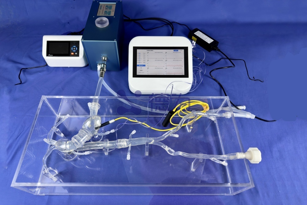
Pressure Visualization Based on a Built-in Sensor Array
A capacitive pressure-sensor array embedding model was designed to target key anatomical landmarks. The sensing unit consists of an ionic hydrogel electrode and a silicon-based dielectric layer; upon compression, deformation of the dielectric layer alters the device’s capacitance. The model incorporates horizontal and vertical electrode channels, which are filled with conductive hydrogel and subsequently cured via UV irradiation, thereby forming discrete sensing units at the channel intersections.
Sensor calibration results demonstrate that artificial valves of different sizes exhibit markedly distinct pressure distributions across key regions of the aorta depending on the implantation depth. For the 29-mm valve, the pressure values at a moderate implantation depth are consistent with the threshold for conduit obstruction risk identified in numerical simulation studies, and this prediction is fully corroborated by the patients’ postoperative outcomes.
Xi’an Dewei Medical Deeply Integrates Sensor Technology with Simulation Models Its accompanying sensors, pressure monitors, and other equipment enable precise testing of medical device operating conditions, complementing the pressure visualization technology developed in this study and providing comprehensive quantitative data to support medical device research and development.

3D-printed aortic root model with an internal sensor array and visualization of pressure distribution after valve implantation.
Discussion
The 3D-printed aortic root model with integrated sensors developed in this study can address current gaps in TAVR clinical practice, helping to optimize strategies for prosthetic valve selection and implantation depth and thereby reducing the risk of postoperative complications. Moreover, the applications of this model can be extended to endovascular embolization, coronary angioplasty, and other fields, thereby overcoming the limited surgical visualization inherent in minimally invasive procedures.
Against this backdrop, Xi’an Dewei Medical’s specialized solutions have demonstrated significant practical value. By leveraging highly transparent soft silicone materials to construct simulation models that accurately replicate human anatomical structures and physiological characteristics, and by integrating a comprehensive suite of testing equipment to create a closed-loop service spanning model fabrication to data acquisition, these solutions not only meet the performance-testing needs of medical-device R&D but also align with industry standards for end-to-end testing. As such, they provide efficient and reliable technical support for the research, development, and validation of minimally invasive interventional medical devices.
Future research should focus on the development of novel 3D-printing materials to enhance sensor resolution and sensitivity. Leveraging its technical expertise in medical simulation and testing, Xi’an Dewei Medical consistently delivers turnkey solutions, thereby accelerating the clinical translation and industrial upgrading of cutting-edge medical technologies.
Keywords:
3D printing,Simulation model,Minimally Invasive Intervention,Medical devices,Silicone vascular model,TAVR,Cardiovascular,Aortic stenosis
Other news
Request a quote
*Please keep your phone accessible—we'll reach out to you within 24 hours.
PARTNERSHIP
Partnership

Contact us
Address: Wangzuo Qujiang, 3269 Yanxiang Road, Yanta District, Xi'an City
Pre-sales Consultation
Request a quote


