


Endoscopic Intervention Urology System Model – Customizable Simulation Silicone Intervention Model
Release date:
2025-06-25 08:52
Urinary tract stones are a common condition in urology. As early as 1901, stones were discovered in the pelvic bones of mummies from ancient Egyptian tombs. Today, they account for 6% to 10% of all urinary system disorders, and their incidence has been steadily rising in recent years, placing a significant burden on healthcare systems worldwide.
Surgical Technique History
Kidney stones (calculus of the kidney) are a common condition of the urinary system, referring to solid formations that develop in the renal calices, renal pelvis, or at the junction where the renal pelvis meets the ureter. In healthy individuals, certain substances normally dissolved in urine can precipitate and accumulate within the kidneys due to various factors, gradually growing over time to form stones—most of which typically remain within the renal pelvis and calyces. The kidneys are the primary site where urinary system stones form; in fact, stones originating in any other part of the urinary tract can initially develop in the kidneys. Ureteral stones typically originate in the kidneys and then travel down the ureters with the flow of urine, leading to symptoms such as ureteral obstruction, severe pain, and hematuria. If left untreated, these conditions can result in serious complications like urinary tract infections and impaired kidney function. Moreover, kidney stones pose a higher risk of directly damaging the kidneys compared to stones forming in other areas of the urinary tract. This underscores the critical importance of early diagnosis and treatment.
Compared to shock wave lithotripsy (SWL), the rapid advancement of ureteroscopy has made treatment possible for most types of ureteral stones—particularly those requiring stent placement prior to SWL (larger than 1.5 cm) or stones that are too large for SWL itself (over 2 cm). Additionally, when the stone is in a favorable position for visualization, ureteroscopy can also be selectively used for renal pelvic stones.
The success of ureteroscopic surgery hinges on successful scope insertion, and for cases where intubation proves challenging, mastering the insertion technique is critical to improving success rates and minimizing postoperative complications. Industry consensus holds that ureteroscopes ≤8F in diameter are considered safe, and ongoing advancements in designing even narrower working channels are enabling more patients to benefit from endoscopic treatment. Meanwhile, guidewires must meet specific requirements: a flexible tip, low friction, and a rigid shaft. Guidewires are often coated with polytetrafluoroethylene (PTFE) or hydrophilic polymers, and they must remain moist before insertion—this not only facilitates smooth placement but also helps safeguard the endoscope’s working channel. As a result, both the design of the instruments and the surgical techniques demand exceptional precision and expertise.
Product Structure

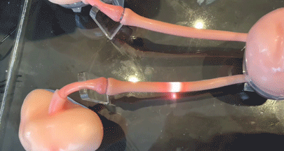
The model can visualize intra-cavity inflammation, stones, and other lesions, and it also allows for the placement of stones to perform lithotripsy procedures.
Endoscopic Intervention Urology System Model It is suitable for both cystoscopy and ureteroscopy. The cystoscope consists of components such as a sheath, obturator, viewing lens, and operational parts. Its slender, flexible body can bend or remain partially rigid, allowing it to easily adapt to various angles required for bladder visualization. Ureteroscopes are categorized into rigid and flexible types. Rigid ureteroscopes are easier to maneuver when entering the ureter, providing clear views of the ureteral lumen. In contrast, flexible ureteroscopes offer greater flexibility and curvature, enabling access to higher segments of the ureter and even the renal pelvis—making them particularly advantageous for exploring curved urinary tract structures.
The urological endoscope model’s overall dimensions perfectly match real human anatomy. The urethral length has been precisely adjusted to ensure the instrument retains flexible maneuverability when entering the ureter from within the bladder. Additionally, the entry point has been moved to the exterior of the housing and incorporates a sliding-slot design. The bevel angle at the junction between the ureter and bladder has been increased, making it easier for the instrument to navigate into the ureter and enhancing user convenience. The ureter is equipped with a quick-connect fitting, enabling effortless disassembly without compromising fluid passage. Meanwhile, an additional outward-facing pathway has been added at the renal pelvis’ distal end, allowing users to insert simulated kidney stones directly into the renal pelvis. This feature also facilitates air evacuation during water injection, ensuring the renal pelvis is fully filled with liquid. Finally, the model boasts a lifelike human-body-inspired casing that not only enhances its aesthetic appeal but also optimizes the anatomical structure to better align with the proportions of a typical male body. To further elevate realism, the model even includes lifelike simulated genitalia.
Product Features
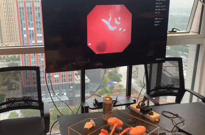
Demonstration of interventional device manipulation within a urological endoscopy model
The instrument moves smoothly through the model, with almost no resistance.
The endoscopic intervention model of the urinary system can be used for Simulation-based procedures such as cystoscopy, ureteroscopy, percutaneous nephroscopy, and transurethral surgery By using this product, you can gain a detailed understanding of the urinary system's anatomical structures and accurately identify key anatomical landmarks. Additionally, it allows you to observe the internal mucosal appearance of the bladder, as well as detect any abnormalities such as inflammation, tumors, or stones. The device also enables biopsies of small lesions within the bladder, helping to establish a definitive pathological diagnosis. Furthermore, it supports simple therapeutic procedures inside the bladder, such as removing minor foreign objects or performing transurethral resection for superficial bladder tumors. Moreover, the instrument is highly effective in diagnosing and treating ureteral conditions—such as ureteral stones—by employing techniques like laser lithotripsy via ureteroscopy to break up and remove the stones. For space-occupying lesions in the ureter, biopsies can be performed to determine their exact nature, while early-stage lesions may even be excised directly using ureteroscopic tools.
How to operate
This model uses a method similar to that of a bronchoscope: before testing with the scope, you need to spray atomized silicone oil onto the inner surface of the model’s lumen. The purpose is to ensure the endoscope moves smoothly and freely through the tube.
The model used for instruments like the Urological Zebra Guidewire is made of transparent silicone (matching the organ structure of this particular model). The transparency of the silicone allows for clearer visualization of how the guidewire instrument moves dynamically through the model.
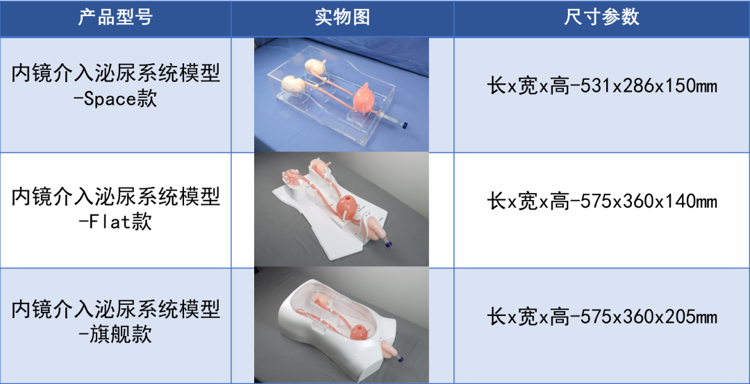
Product Configuration Table
Keywords:
Urology,Urinary tract stones,Kidney stones,Cystoscopy,Ureteroscopy,Percutaneous Nephroscopy,Transurethral surgery,Silicone Vascular Model,3D printing,Medical simulation,Equipment Development,Minimally Invasive Interventional Testing,Medical device testing
Other news
Request a quote
*Please keep your phone accessible—we'll reach out to you within 24 hours.
PARTNERSHIP
Partnership

Contact us
Address: Wangzuo Qujiang, 3269 Yanxiang Road, Yanta District, Xi'an City
Pre-sales Consultation
Request a quote


