


DeWei All-Brain Silicone Vascular Model: Technology Empowers DSA Angiography Research
Release date:
2025-06-26
Cerebrovascular diseases significantly contribute to global mortality and long-term disability (Vaduganathan et al., 2022). In recent years, thanks to advances in medical and imaging technologies, the mortality rates associated with intracranial arterial (IA) diseases—such as intracranial artery stenosis (ICAS), middle cerebral artery occlusion (MCAO), and intracranial aneurysms—have declined (Hess, 2018).
DeWei All-Brain Silicone Vascular Model: Technology Empowers DSA Angiography Research
Cerebrovascular diseases significantly contribute to global mortality and long-term disability (Vaduganathan et al., 2022). In recent years, advances in medical and imaging technologies have led to a decline in mortality rates associated with intracranial arterial (IA) conditions such as intracranial artery stenosis (ICAS), middle cerebral artery occlusion (MCAO), and intracranial aneurysms (Hess, 2018). Among these technologies, digital subtraction angiography (DSA) stands out as an imaging technique that captures a series of images to visualize the flow of contrast agents through blood vessels—typically over a period of 3 to 15 seconds, with a frame rate ranging from 3 to 5 frames per second (Jin et al., 2020). By subtracting pre-contrast images, DSA effectively removes background structures like bone, enhancing the visibility of vessels filled with contrast. Thanks to its inherent superior spatial and temporal resolution, DSA can accurately reveal intricate details of vascular lesions—details that may remain unclear with alternative imaging methods such as computed tomography angiography (CTA) or magnetic resonance angiography (MRA), which sometimes fall short in providing definitive diagnoses (Hess, 2018). As a result, DSA is widely regarded as the gold standard for studying the structure of diseased vessels, interpreting arterial blood flow dynamics, and guiding endovascular interventions (Shaban et al., 2022).
[Figure 1. Anterior and Lateral DSA Images of the Cranioencephalic Region]
As silicone vascular printing technology continues to advance, With the help of sophisticated vascular models, it is possible to obtain richer and more multidimensional insights into the spatial structure of blood vessels as well as hemodynamic information. , which is of great significance for deepening the understanding of cerebrovascular diseases and guiding clinical diagnosis and treatment decisions.
Facing this demand, DeWei Medical has continuously explored and developed a comprehensive brain silicone vascular model specifically designed for DSA image testing. The product includes a 3D-printed, full-brain silicone vascular model, an injection pump, and a circulation pump. Crafted with high-tech materials, it accurately replicates the intricate structure of cerebral blood vessels at a 1:1 scale, faithfully recreating even the tiniest vascular details. Additionally, it features lesion-specific vascular segments tailored for interventional procedures, with unilateral inclusion of aneurysms and stenotic lesions to enhance realism. The model also mimics the dynamic internal environment of the brain, complete with an external circulation pump that ensures fluid flow through every cerebral vessel branch. This innovative tool is versatile enough to support a wide range of applications, including consumable product testing, coil embolization procedures, and thrombectomy surgeries—significantly boosting testing efficiency while cutting down on R&D costs.
In cerebral vascular DSA diagnostics, the injection rate and distribution pattern of the contrast agent directly affect image quality and diagnostic accuracy. Clinical studies have shown that when the contrast agent is injected at a rate slower than 5 ml/s, vascular visualization becomes inadequate, increasing the risk of missing subtle lesions. Conversely, injecting too quickly—exceeding 8 ml/s—can lead to turbulence, resulting in image artifacts. Moreover, the concentration gradient distribution of the contrast agent within the vessels must closely mimic the body’s natural hemodynamics to accurately reproduce vascular images reflecting the true pathological conditions.
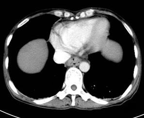
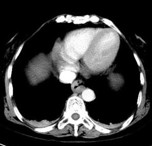
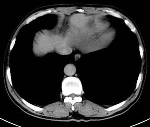
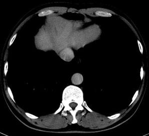
Contrast agent injection rates of 4.0 mL/s, 3.5 mL/s, 3 mL/s, and 2.5 mL/s—CTV images in axial views at the inferior vena cava-right atrium junction for each experimental group.
The DSA whole-brain silicone vascular model addresses the challenges of contrast agent application, achieving three major technological breakthroughs:
- Precise flow rate control: The high-precision injection pump supports a wide range of flow rate adjustments from 0.1 to 8 ml/s. Measured at standard injection speeds, the time it takes for the contrast agent to uniformly fill the unilateral cerebral vascular region closely matches the clinically observed imaging time window.
- Dynamic concentration simulation: Through the coordinated control of the circulation pump and the infusion pump, the model can simulate the dilution process of contrast agents within blood vessels. Testing has shown that, at a circulation flow rate of 11 L/min, the contrast agent concentration decay curve closely matches real-world human data, accurately replicating the entire process of contrast agent diffusion and metabolism as it moves with blood flow.
- Image-level detail presentation: A 1:1 replica of the cerebral vascular structure (with an error margin of ≤0.1 mm) ensures that the flow path of the contrast agent within the tiny branch vessels closely matches its natural course in the human body. In DSA imaging tests, secondary branch vessels such as the lenticulostriate arteries and anterior choroidal arteries demonstrated clear visualization, fully meeting clinical requirements for observing even the most subtle lesions.
Precise flow simulation, replicating the human body's real circulation
To match the real blood flow in human cerebral vessels, the model is equipped with a high-precision injection pump that plays a dual and critical role. On one hand, its advanced mechanical and algorithmic control ensures stable flow rates, faithfully replicating the dynamic characteristics of fluid movement within vascular branches—whether it’s the pulsatile nature of arterial blood flow or the smooth, steady state of venous return. This precision not only provides reliable data for DSA imaging tests but also enables realistic surgical simulations.
On the other hand, rational use of contrast agents is crucial during DSA examinations. Clinical studies show that when the rate of contrast agent administration exceeds the safety threshold within a given time frame, patients experience a significantly increased risk of developing contrast-induced nephropathy, allergic reactions, and other signs of toxicity. This infusion pump supports precise flow rate adjustment from 0.1 to 5 ml/min, enabling real-time, dynamic control of the water-to-contrast agent ratio delivered into the bloodstream—ensuring a perfect balance. Simulation tests have demonstrated that, under standard imaging procedures, the pump’s accurate regulation keeps the contrast agent concentration within the model consistently stable, with fluctuations controlled within ±3%. This effectively prevents potential side effects caused by contrast agent buildup, helping medical staff master safe contrast agent usage strategies during simulation training and ultimately reducing risks in actual clinical procedures.
- Intelligent hydration system ensures long-lasting circulation
To address the potential issue of water tank level drop caused by long-distance control of the infusion pump, the R&D team has innovatively designed an automatic water-refilling device. This device continuously monitors the water tank’s liquid level in real time and automatically activates a refill pump to draw water from the storage unit whenever the level falls below a preset threshold—ensuring the circulation system can operate sustainably for up to 1 hour. Additionally, the system can be customized according to customer needs to offer extended endurance options, perfectly meeting the demanding requirements of prolonged surgical simulations and complex experiments.
- Independent wastewater recycling system eliminates cross-contamination.
To prevent backflow of discharged wastewater from contaminating the original water tank, the model features an externally connected wastewater collection tank design. During experiments, used fluids are directly drained into the separate wastewater tank, where a one-way valve and piping system ensure zero backflow—effectively maintaining the purity of the original water tank and guaranteeing the accuracy and reliability of each simulation test.

Keywords:
Cerebral blood vessels,Imaging technology
Request a quote
*Please keep your phone accessible—we'll reach out to you within 24 hours.
PARTNERSHIP
Partnership

Contact us
Address: Wangzuo Qujiang, 3269 Yanxiang Road, Yanta District, Xi'an City
Pre-sales Consultation
Request a quote


