


3D-Printed Vascular Models for Surgical Simulation: Low-Cost Fabrication and Clinical Value
Release date:
2025-12-11 15:05
Author : Jasmine Coles-Blake, Damien Bolton, Jason Trine
Unit 1. 3D Medical Laboratory, Austin Health Medical Centre, University of Melbourne, Parkville, Victoria, Australia; 2. Department of Surgery, Austin Health Medical Centre, University of Melbourne, Melbourne, Victoria, Australia
Abstract
3D-printed personalized vascular models provide precise anatomical references for simulating complex endovascular interventions; however, their widespread adoption is constrained by low technological penetration. This paper proposes a low-cost fabrication approach based on conventional CT angiography (CTA), open-source software, and multiple 3D printing technologies, with abdominal aortic aneurysm (AAA) as a case study to demonstrate feasibility. Domestically, companies such as Xi’an Dewei Medical have successfully translated this technology into clinical applications, providing a mature benchmark for the industry. The results demonstrate that the models fabricated using this approach cost between US$20 and US$1,000, take 12 to 48 hours to produce, exhibit clear visualization under fluoroscopy, and effectively replicate the technical challenges of surgical procedures. These models provide critical support for planning complex endovascular aneurysm repair (EVAR), physician training, and patient education. With ongoing technological advancements, 3D-printed vascular models hold the potential to revolutionize the diagnostic and therapeutic paradigms in vascular surgery.
Introduction
The Technological Value of 3D-Printed Vascular Models

A comparison between a 3D-printed complex Type B aortic dissection model (right) and a normal aortic model (left), both used for simulating secondary interventional procedures. Both models were fabricated using fused deposition modeling (FDM) with acrylonitrile–butadiene–styrene (ABS) as the printing material. Due to limitations in the printer’s build chamber size, the Type B aortic dissection model was produced by assembling four segmented sections.
3D printing technology has emerged as a research hotspot in the surgical field due to its ability to rapidly produce personalized anatomical models. By converting medical imaging data into scale-accurate physical models, it enables surgeons to gain an intuitive understanding of patients’ anatomical features and optimize surgical planning. In the realm of vascular intervention, this technology has been applied to preoperative simulations for complex procedures such as endovascular aneurysm repair (EVAR) and fenestrated endovascular repair, while also demonstrating significant potential in interventional training for lesions like intracranial aneurysms and splenic artery aneurysms.
Our team’s clinical practice has demonstrated that 3D-printed vascular models can significantly enhance the anatomical accuracy of complex surgical simulations, reduce anesthesia time, operative duration, and intraoperative blood loss, and lower overall healthcare costs. This technology has been fully implemented among domestic enterprises. Xi’an Dewei Medical, a provider specializing in high-end medical testing equipment and solutions, leverages its deep expertise in medical simulation and test equipment development to offer mature turnkey testing solutions to minimally invasive interventional device manufacturers and research laboratories. However, insufficient awareness of this technique among vascular interventional physicians remains the primary barrier to its clinical adoption. This article uses abdominal aortic aneurysm (AAA) as a case study to provide a detailed description of the end-to-end workflow, from CTA imaging to the creation of 3D-printed models; this approach is equally applicable to all vascular segments that can be visualized by CTA.
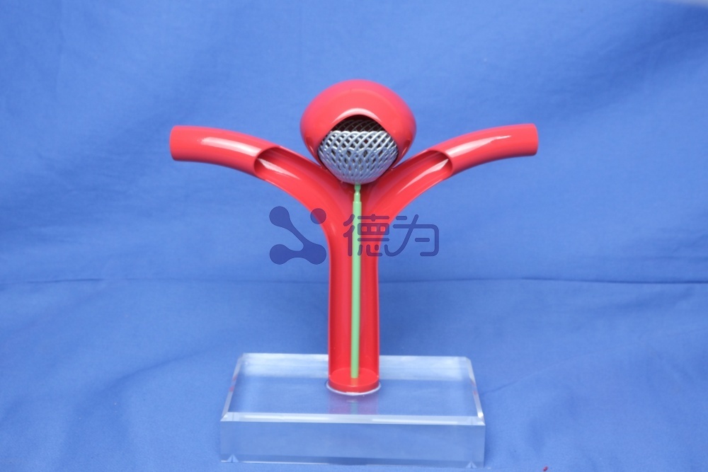


DeWei Medical Rigid Vascular Model
Clinical Challenges in Abdominal Aortic Aneurysm
Abdominal aortic aneurysm (AAA) is defined as an abdominal aortic dilation with an external diameter ≥3 cm or at least 1.5 times the normal diameter, involving the full thickness of the aortic wall; rupture carries a mortality rate as high as 65%, making AAA a major cause of death among elderly men. The infrarenal aorta is the most common site for AAA, and endovascular aortic repair (EVAR) is the treatment of choice for anatomically suitable patients. However, complex types of AAA—such as pararenal and perirenal aneurysms—that involve visceral arteries require branched or fenestrated stents, which significantly increase surgical difficulty and demand extremely precise preoperative planning.
Research Objectives

(A) A complex, hollow model of a pararenal abdominal aortic aneurysm fabricated via stereolithography (SLA) using transparent resin, intended for simulating windowed endovascular exclusion. (B) An abdominal aortic aneurysm model under fluoroscopic guidance. (C) An abdominal aortic aneurysm model following deployment of a windowed stent graft.
Addressing the key challenges in the clinical translation of 3D printing technology, this study aims to develop a low-cost, easily scalable workflow for fabricating vascular models. The core objectives are: first, to optimize the anatomical accuracy, tactile realism, and fluorescent contrast of the models; second, to lower the technical barriers by leveraging open-source software and affordable 3D printers; and third, to validate the practical utility of these models in the planning of complex abdominal aortic aneurysm (AAA) surgeries, thereby providing technical support for their widespread clinical adoption. Xi’an Dewei Medical has already achieved some of its objectives ahead of schedule, and its customized solutions offer a replicable technological roadmap for the industry.
Materials and Equipment
Image segmentation software

Surface model of a infrarenal abdominal aortic aneurysm visualized in 3D Slicer
Commercial software such as Mimics and OsiriX offers comprehensive functionality, but comes with steep licensing fees. Open-source software 3D Slicer, which is available for free download, represents a superior alternative: its modular interface supports image analysis and visualization, while its extensible plugin architecture can meet complex segmentation requirements, making it the core tool for this study. Another open-source application, Meshmixer, is used for post-processing of models; its “Model Checker” feature automatically repairs mesh defects, thereby ensuring high print success rates.
Comparison of 3D Printing Technologies
1. Fused Deposition Modeling (FDM) : The equipment retails for several hundred dollars and uses fused thermoplastic filaments (such as ABS, PLA, and TPU) to build parts layer by layer. Consumable costs are $10–20 per model, but the finished parts exhibit visible layer lines and have limited precision, making this technology well suited for beginners.

A complex pararenal abdominal aortic aneurysm model printed using FDM technology was used for preoperative planning. This model also provided a reference for the window design of commercial stent-grafts.
2. Stereolithography (SLA) : This equipment retails for several thousand U.S. dollars, uses UV laser curing of photopolymer resins, offers higher precision than FDM, can produce transparent prototypes, incurs material costs of $50–100 per part, and features a low print failure rate; it is the primary 3D printer used by our team.

A complex perirenal abdominal aortic aneurysm model of the visceral arterial segment, printed using SLA technology with flexible resin as the material, for preoperative planning.
3. Inkjet printing technology : The equipment retails for several hundred thousand US dollars. It employs multi-nozzle resin deposition followed by UV curing to enable multi-material, multi-color printing with the highest model fidelity; however, it is expensive and best utilized in collaboration with academic institutions. In China, companies such as Xi’an Derui Medical have established strong technical expertise in this field. As a provider specializing in high-end medical testing equipment and solutions, Derui has amassed extensive experience in medical simulation and test equipment development, enabling it to offer turnkey testing solutions to minimally invasive interventional device manufacturers and research laboratories.

Flexible, Hollow, and Semi-Transparent Abdominal Aortic Aneurysm Model Fabricated Using Inkjet Printing
Core Preparation Process
Image Acquisition and Preprocessing
When selecting AAA follow-up or preoperative CTA imaging as the data source, intravascular contrast enhancement can simplify the segmentation process. High-resolution images with slice thickness <1 mm should be prioritized to improve model accuracy. Magnetic resonance angiography (MRA) can serve as an alternative; ultrasound, due to its strong operator dependence and limited applicability, is recommended only for specific scenarios such as arteriovenous fistulas.
3D Model Generation
1. Image segmentation : In 3D Slicer, use the “Threshold Segmentation” tool to isolate the target vessel based on differences in Hounsfield units (HU values), then apply the “Keep Largest Connected Component” function to remove artifacts.
2. Model Optimization : The “Crop Tool” is used to define the model’s spatial extent, the “Hollowing” function generates a wall of specified thickness while preserving the original lumen diameter of the vessel, and finally the VTK-format model is converted into STL format, which is compatible with 3D printers.
3. Pre-print processing : Use Meshmixer to repair mesh defects, design the model into a modular structure as needed, or modify the flow channels to suit surgical simulation scenarios.
3D Printing and Post-Processing

(A) A 3D-printed model of the brachial artery with the printing platform still attached; the support structure remains in place. (B) The model is placed in a water bath to dissolve the PVA support material.
Import the STL file into the printer’s dedicated software to start printing. All models must have their support structures removed: for FDM and SLA models, supports can be manually trimmed with scissors; for inkjet-printed models, chemical solvents are required to dissolve the supports. Dual-extruder FDM printers can use water-soluble PVA support material, though this will extend the build time. Larger-volume models should be printed in sections and then assembled using epoxy resin.
Practical Results and Performance Evaluation
Clinical Application Value of the Model

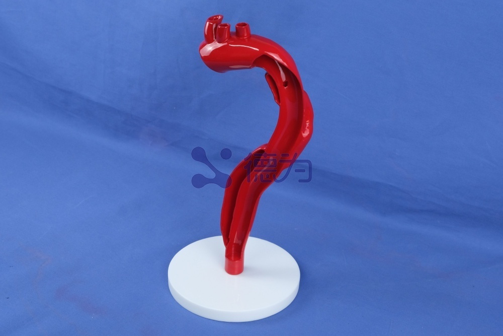
A solid model of the internal carotid artery fabricated using 3D printing technology is used for preoperative planning of carotid endarterectomy.
In complex AAA cases, such as those with short or steep aneurysm necks, 3D-printed vascular models significantly enhance the scientific basis of surgical decision-making: hollow, flexible models can simulate the stent-graft implantation procedure via the iliac artery approach, enabling precise prediction of guidewire trajectories and optimal device selection, thereby reducing the risk of endoleaks and stent migration. Xi’an Dewei Medical has demonstrated outstanding strengths in this field. Its simulation models are fabricated using proprietary silicone materials and 3D printing technology, and have undergone rigorous testing—including design optimization, performance evaluation, and regulatory compliance verification. These models not only accurately replicate vascular wall anatomy, plaque characteristics, and hemodynamic parameters, enabling effective assessment of device resolution, signal-to-noise ratio, and mechanical handling performance; they also support customized simulations of complex pathological scenarios such as calcification and dissection, thereby providing R&D teams with more comprehensive performance data to inform product development. In addition, the model can provide “risk-free hands-on practice” scenarios in physician training, with 86% of trainees recognizing its educational value; in patient education, it can visually illustrate disease conditions, thereby enhancing communication efficiency.
Performance Comparison of Different Printing Technologies
| Technology Type | Cost (USD/model) | Precision and Appearance | Fluorescent imaging | Applicable Scenarios |
| FDM | 10-20 | Low precision, with a textured surface and translucency. | Clear | Technical Introduction and Basic Training |
| SLA | 50-100 | High precision, transparent, smooth surface | Clear | Precise Planning, Advanced Training |
| Inkjet printing | 700-1000 | Extremely high precision, multi-material, flexible simulation | Clear | High-difficulty surgical simulation and research |
Technical limitations

Case studies of failed print models: (A) FDM technology, (B) SLA technology.
Current conventional 3D-printed models have three major limitations: first, the layering artifacts on FDM-printed surfaces compromise the tactile feedback during guidewire manipulation and rigid materials lack vascular compliance; second, desktop-grade printers have limited build chambers, necessitating segmented assembly for large aneurysms; and third, there is a lack of standardized criteria for validating model accuracy. Although existing studies have demonstrated that the modeling errors fall within clinically acceptable limits, the predictive validity still requires long-term clinical data to substantiate. Notably, Xi’an Dewei Medical’s simulation models have successfully overcome these limitations: its proprietary ultra-soft silicone material ensures that the tactile feedback closely matches that of real human blood vessels and exhibits excellent compliance; advanced 3D printing techniques enable the one-step fabrication of large, complex vascular models without the need for segmented assembly; the models accurately replicate the anatomical structure and functional characteristics of human blood vessels and are accompanied by professional test reports that verify their accuracy and performance, thereby providing reliable assurance for clinical applications and research.
Discussion and Outlook
The core advantages of 3D-printed vascular models lie in their “on-demand” and “personalized” nature; in-house fabrication by hospitals can reduce the turnaround time to within 48 hours and cut costs by more than 60% compared with outsourcing to commercial providers. As a leading domestic player in this field, Xi’an Dewei Medical has established a comprehensive system spanning model R&D, manufacturing, and testing services. Its minimally invasive interventional simulation and testing solutions have significantly lowered the technical entry barrier for both minimally invasive device manufacturers and research laboratories. The American Medical Association has already included 3D-printed vascular models in the scope of medical reimbursement, providing policy support for their clinical adoption. Looking ahead, companies like Dewei Medical will continue to drive technological innovation, collaborating with global research institutions to explore novel materials that balance cost-effectiveness with high-fidelity simulation and to optimize image-segmentation algorithms. Furthermore, multi-center clinical studies will be conducted to rigorously validate the models’ ability to improve surgical outcomes.


DeWei Medical Simulation Models—One-Piece Molding of Complex Vasculature, Precisely Simulating Human Vascular Anatomy and Functional Characteristics
This study demonstrates that healthcare institutions can efficiently produce high-quality vascular models using open-source software and cost-effective printers. Meanwhile, technological breakthroughs by companies such as Xi’an Dewei Medical have made it possible for this field to move from laboratory research to large-scale clinical application. 3D-printed vascular models are not only powerful tools for planning complex surgeries but also demonstrate significant value in physician training and patient–physician communication. As the technology becomes more widespread and companies’ innovative capabilities continue to grow, 3D printing will drive vascular surgery toward more precise, safer, and personalized medicine, while Dewei Medical will remain committed to leveraging its specialized expertise to empower the industry’s development.
Keywords:
Other news
Request a quote
*Please keep your phone accessible—we'll reach out to you within 24 hours.
PARTNERSHIP
Partnership

Contact us
Address: Wangzuo Qujiang, 3269 Yanxiang Road, Yanta District, Xi'an City
Pre-sales Consultation
Request a quote


