


The Application Value of 3D-Printed Individualized Aortic Arch Models in Medical Device Testing and R&D Validation
Release date:
2026-01-14 17:02
Authors: Smruti Mahapatra, Vishal N. Bimalasetty, Abdul Rahim, et al.
Abstract
Research Background
Cerebrovascular diseases are a leading cause of death and disability worldwide, with endovascular therapy serving as the cornerstone of treatment for ischemic stroke. Anatomical abnormalities such as a tortuous aortic arch frequently impede the navigation of interventional devices and limit their performance, thereby constituting a critical bottleneck for device optimization and advancement.
Research Methods
Based on imaging data from four patients with complex vascular anatomy, 3D-printed models of the aortic arch and its branch vessels were fabricated to establish a personalized vascular anatomical simulation platform for the testing and evaluation of interventional devices.
Research Results
3D-printed models can accurately replicate the anatomical details of blood vessels, with measured vessel diameters in close agreement with reported literature values, thereby confirming their anatomical accuracy. On average, each model requires 4 hours of digital processing and 13.71 hours of printing, with material costs of approximately US$17.31, indicating potential for low-cost, large-scale fabrication and making them suitable as standardized platforms for device testing.
Research Conclusions
This model provides a highly realistic in vitro platform for the testing and R&D validation of interventional medical devices, enabling effective assessment of device fit and procedural safety, providing data-driven support for device optimization and iteration, and enhancing the device’s ability to adapt to complex anatomical scenarios.
Keywords : Anatomical variation; cerebrovascular disease; 3D printing; patient-specific models; interventional medical devices; device testing; R&D validation
Introduction
Cerebrovascular diseases, characterized by high mortality and disability rates, impose stringent performance requirements on interventional medical devices. As stroke ranks as the second leading cause of death worldwide and its prevalence is increasing among younger populations, this trend further drives the need for the development of highly effective and safe interventional devices⁽¹⁾⁽²⁾⁽³⁾. The aortic arch, serving as a central access route for interventional procedures, is subject to anatomical variations—particularly Type III aortic arches and vascular tortuosity—that can impede device delivery, reduce navigation accuracy, and even lead to vascular injury⁽⁹⁾⁽¹⁰⁾.
Conventional imaging modalities such as CTA and MRA are unable to generate physical simulation environments, making it difficult to accurately assess the mechanical compatibility of devices during dynamic interventions—this has become a major bottleneck in research and development validation. In contrast, 3D printing technology, with its high-fidelity and patient-specific advantages, can construct physical vascular models based on patient imaging data and, through careful material selection, mimic vascular mechanical properties, thereby providing a visual platform for performance testing and optimization of catheters, guidewires, and other interventional devices⁽¹¹⁻¹⁵⁾. This study fabricated models from four complex vascular cases to explore their clinical value in device testing and R&D.
Clinical History
This section presents four cases of patients with aortic arch anatomical anomalies (Figure 1), in which vascular tortuosity limits the use of conventional interventional devices, thereby providing representative scenarios for device compatibility testing. Cases 1–3 underwent CTA, while Case 4 underwent MRA due to iodine allergy.
Case 1 (90 years old) and Case 3 (82 years old) both had severe, tortuous Type III aortic arches, which prevented multiple guidewires and catheters from safely reaching the target site. Instrument manipulation was markedly constrained by the anatomical anatomy, resulting in failure of the interventional procedure. Case 2 (76 years old) had bilateral vertebral artery tortuosity that obstructed catheter-based navigation; despite changing instruments and access routes, effective recanalization was still not achieved, highlighting significant issues with instrument compatibility. In Case 4 (79 years old), instrument delivery via the femoral artery was impeded, but successful recanalization was achieved after switching to radial artery access, suggesting that this model can be used to evaluate differences in instrument compatibility across different access routes.

Figure 1: Coronal DICOM images of the aortic arch in four patients, all demonstrating anatomical abnormalities accompanied by vascular tortuosity, thereby providing a complex anatomical simulation scenario for device testing (A: Case 1; B: Case 2; C: Case 3; D: Case 4).
Research Methods
Case Selection and Ethical Approval
Four of the aforementioned cases were selected to create anatomical models, which accurately replicate the complex and tortuous anatomical features encountered in high-difficulty clinical device procedures. This study was approved by the Oxena Clinical Foundation’s Institutional Review Board (approval number: STUDY00000589) and complies with the relevant provisions of the Declaration of Helsinki.
3D Model Creation (Tailored to Medical Device Testing Requirements)
Patient DICOM-format images were acquired and imported into Vertex software, after which vascular model segmentation was performed using the Mimics 26 suite to generate stereolithography files. The solid model was then post-processed into a 1-mm-thick hollow structure to simulate the real vascular wall for device passage testing. The models were fabricated using Formlabs 80A flexible resin and a Form 3B+ printer; the resin’s flexibility accurately mimics the fundamental mechanical properties of blood vessels. Following isopropyl alcohol washing and 60°C UV curing, the final models retained the complete vascular branching architecture (Figure 2).

Figure 2: Workflow for 3D-printed model fabrication: image processing and segmentation → generation of stereolithography files → printing of hollow models using flexible resin, to meet the requirements for instrument passage and push-through testing.
Research Results
The 3D model and the printed prototype (Figures 3 and 4) accurately reproduce the tortuosity of the type III aortic arch and the branching angles, providing a precise simulation environment for evaluating device navigation performance. Vascular diameter measurements (see Table) show that the mean diameters of the ascending aortic trunk and the descending aortic trunk are 33.64 mm and 26.90 mm, respectively, while those of the brachiocephalic trunk, the subclavian artery, the common carotid artery, and the vertebral artery are 14.22 mm, 7.57 mm, 7.92 mm, and 4.73 mm, respectively. These measured values are in excellent agreement with reported literature data⁽²¹⁻²⁷⁾, thereby meeting the requirements for device size compatibility testing. The application of such high-fidelity models also aligns with the current demand in the medical-device testing field for specialized simulation platforms, providing a foundational support for clinical translation.
This model offers the advantages of low cost and rapid prototyping, enabling the mass production of samples representing various anatomical types to build a device-testing database and providing efficient support for R&D iteration.

Figure 3: Coronal (top row) and sagittal (bottom row) views of the 3D model of the aortic arch, providing precise morphological reference for device compatibility testing.

Figure 4: A flexible resin 3D-printed model that can simulate vascular compliance and is suitable for device manipulation testing.

Table: Diameters of Major Vessels in the 3D-Printed Model (Unit: mm)
Discussion
3D-printed patient-specific models provide a high-fidelity, reproducible in vitro testing platform for interventional devices, thereby addressing the limitations of conventional imaging techniques. These models not only accurately replicate vascular anatomical parameters, but also use flexible resin materials to simulate the mechanical response of the vessel wall, enabling effective evaluation of critical performance metrics such as device delivery resistance and navigation accuracy, and providing direct data support for structural optimization. In this field, Xi’an Dewei Medical has established a strong technical expertise. As a supplier specializing in high-end medical testing equipment and solutions, the company offers simulation models designed for medical device engineers that achieve a 1:1 reproduction of human anatomical structures, enabling precise alignment with complex vascular testing requirements.
Models fabricated based on four cases of Type III aortic arch anomalies can simulate highly challenging clinical procedural environments, enabling targeted evaluation of the compatibility of devices under different design schemes and thereby shortening the clinical translation cycle for medical devices. Compared with generic testing models, these patient-specific models can accurately replicate individual anatomical features, precisely reveal device compatibility shortcomings, and—thanks to their low cost and rapid prototyping capabilities—facilitate the establishment of a standardized test library for systematic performance validation of medical devices. Meanwhile, DeWei Medical has further upgraded its simulation technology. Its vascular models are made from a proprietary, highly transparent soft silicone material that overcomes the limitations of traditional materials, enabling precise reproduction of human hemodynamics and vascular physiological characteristics. When paired with integrated equipment such as sensors, flow monitors, pressure monitors, and push-force testing devices, these models allow for multidimensional, high-precision quantitative testing of medical device performance, thereby providing more comprehensive data support for device R&D.
DeWei Medical Neurointerventional Vascular Model
This study has several limitations: the model does not fully replicate vascular elasticity and hemodynamic characteristics; the case cohort is limited in scope; printing resolution and material properties are constrained by current technological limitations; and further optimization is needed to enhance testing accuracy. However, Leveraging its extensive expertise in medical simulation, testing equipment, medical software development, and test-system integration, DeWei Medical has developed mature technical solutions that effectively address the technological gaps in such research and clinical trials. The company provides turnkey testing solutions for minimally invasive interventional device manufacturers and high-end research-oriented medical laboratories, covering the entire process—from model development to precision testing.
Future Research Directions
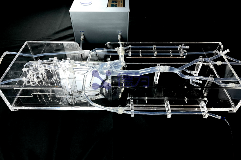
A prototype hemodynamic simulation test platform built on this model (Figure 5) employs peristaltic pumps to create a fluid circulation system, enabling the simulation of blood flow effects on device operation; subsequent process optimization will further reduce costs. In the future, biomimetic composite materials will be incorporated to precisely replicate vascular mechanical properties; physiological monitoring modules will be integrated to collect quantitative data on device performance; the model library will be expanded to encompass a broader range of scenarios; and finite-element analysis will be combined with experimental testing to enable mutual validation between empirical data and computational simulations, thereby enhancing the scientific rigor of R&D. This development direction is highly aligned with DeWei Medical’s technological strategy; its integrated testing system has achieved seamless integration of hemodynamic simulation, multi-parameter monitoring, and precise quantitative analysis, providing efficient and reliable technical support for the R&D and validation of medical devices and promoting the standardization and precision of testing workflows.

Figure 5: Prototype of the hemodynamic simulation test platform, capable of real-time evaluation of device performance in a dynamic blood flow environment.
Conclusion
Low-cost, high-fidelity 3D-printed individualized aortic arch models serve as an excellent ex vivo platform for the testing and R&D validation of interventional medical devices. They can precisely replicate complex vascular anatomical features, providing clinically relevant scenarios for device compatibility and safety assessments, and supporting the optimization and iterative development of these devices. And Through technological innovation, Dewei Medical has deeply integrated simulation models with integrated testing equipment, achieving both high-fidelity reproduction of anatomical structures and precise quantification of test data. This provides medical device engineers with specialized tools that are more closely aligned with R&D needs. As the technology continues to mature, such specialized testing solutions are poised to become standard configurations in medical device R&D, driving the development of devices that are better suited to complex anatomy and deliver greater safety and efficiency. Dewei Medical will also continue to empower the R&D and scientific innovation of minimally invasive interventional devices through end-to-end solutions.
Keywords:
3D printing,Medical devices,Interventional medical devices,Minimally Invasive Intervention,Simulation model,Medical Device Engineer,Aortic arch,Cerebrovascular,Neurointervention
Other news
Request a quote
*Please keep your phone accessible—we'll reach out to you within 24 hours.
PARTNERSHIP
Partnership

Contact us
Address: Wangzuo Qujiang, 3269 Yanxiang Road, Yanta District, Xi'an City
Pre-sales Consultation
Request a quote


